[ad_1]

When Jedi Knights have to vanquish an enemy, they whip out their trusty lightsabers. Sooner or later, because of Johns Hopkins researchers, docs looking for to crush most cancers could wield minuscule molecular nanoSABERs that permit them to have a look at tumors in methods by no means earlier than potential.
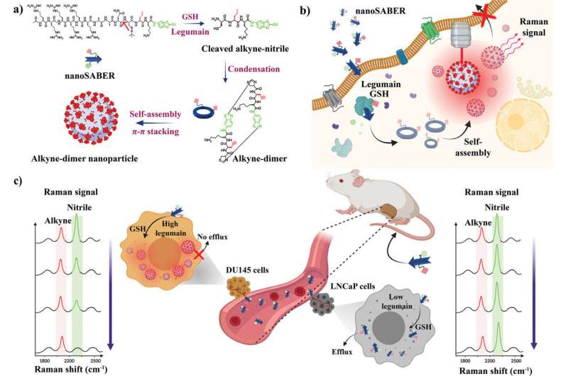
Impressed by the method cells use to assemble proteins, a staff led by two researchers—Ishan Barman on the college’s Whiting College of Engineering and Jeff W. Bulte, a professor of radiology and radiological science on the College of Medication who can be affiliated with JHU’s Institute for NanoBioTechnology—has created infinitesimal probes that mild up once they encounter sure enzymes present in most cancers cells. The flexibility to visualise tumors of their entirety—and early—may considerably improve most cancers imaging, inform therapy choices, and enhance affected person outcomes.
“This might be a recreation changer for most cancers therapy,” mentioned Barman, an affiliate professor of mechanical engineering on the Whiting College, of the self-assembling biorthogonal enzyme recognition (nanoSABER) probes. The staff’s outcomes seem in Superior Science.
At present, tissue biopsies are the gold commonplace for detecting most cancers, although they are often inexact and even miss elements of tumors lurking within the margins. The Johns Hopkins staff’s strategy may remedy that drawback, permitting clinicians to visualise cancerous exercise throughout complete tumors, offering insights into their potential aggressiveness.
Enzymes, particularly legumain, play a number one function within the growth and development of most cancers.
The staff’s new software assembles itself within the presence of those cancer-related enzymes and emits a sign that may then be picked up by Raman spectroscopy, a visualization approach that analyzes molecular vibrations to establish and characterize substances. This enables the probes to pinpoint most cancers cells precisely.
The Johns Hopkins staff says its technique additionally may permit clinicians to extra precisely monitor the buildup of most cancers medicine in tumors throughout therapy, offering a sign of how effectively these remedies are working.
“The probes’ capacity to supply a transparent have a look at the molecular, mobile, and tissue ranges offers a complete perspective,” mentioned lead writer Swati Tanwar, a post-doctoral fellow in mechanical engineering. “It’s crucial to grasp what is de facto taking place on the tumor margins to make sure full most cancers elimination and reduce the possibilities of recurrence.”
Examine co-authors at Johns Hopkins embrace Behnaz Ghaemi, Piyush Raj, Aruna Singh, Lintong Wu, Dian R. Arifin, and Michael T. McMahon. The staff additionally included Yue Yuan of the College of Science and Know-how of China.
Extra data:
Swati Tanwar et al, A Sensible Intracellular Self‐Assembling Bioorthogonal Raman Lively Nanoprobe for Focused Tumor Imaging, Superior Science (2023). DOI: 10.1002/advs.202304164
Offered by
Johns Hopkins College
Quotation:
Researchers develop tiny nanoSABERs to help battle in opposition to most cancers (2023, October 12)
retrieved 12 October 2023
from https://phys.org/information/2023-10-tiny-nanosabers-aid-cancer.html
This doc is topic to copyright. Aside from any honest dealing for the aim of personal examine or analysis, no
half could also be reproduced with out the written permission. The content material is offered for data functions solely.
[ad_2]
